Laser optics offer microscopic views beneath the surface
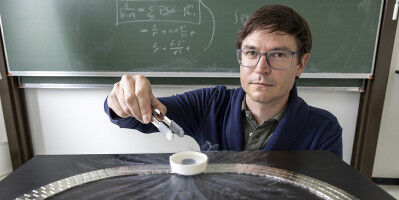
Video of the inside of a fly's eye: Saideh Saghafi is developing laser optics which support high-resolution 3D microscopy. Fine venules, thin branches of nerve tracts - thanks to the ultramicroscope developed at the Bioelectronics Department of the Institute for Solid-State Electronics at the Vienna University of Technology, the tiniest details of biological tissues can be represented in 3D. Laser beams are used to look inside flies, mice, or medical tissue samples. The laser technology and the optics in the device were developed by Saideh Saghafi. Using various optical tricks, she has managed to turn a laser beam into an extremely thin two-dimensional laser surface, which can be shone through samples layer by layer. She has now been awarded a major optics prize for this work. Tissue made transparent - Biological tissue tends to be opaque, with light being scattered at the interfaces between different materials.